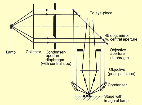
Reflected-light, dark-field illumination with an ellipsoidal ring-mirror reflector and a concave ring-mirror condenser
Figure from "Microscope Photometry", H. Piller, Springer, ISBN 3-540-08094-5
This is the ray path in a epi-illuminated (light from above the sample)
microscope in dark-field mode.
The light is emanating from the lamp to the left. The light is made into a
roughly parallel beam by the Collector. Note that all lenses are only
shown as a line indicating their principal plane.
Most of the light is then blocked by the Condenser-aperture-diaphragm,
which has a central stop that blocks the central rays of the beam.
The light is then reflected by a 45° mirror with a circular hole
in the center.
The light is traveling vertically down
on the outside of the actual objective lens. It is
focused on the sample by an inner mirror at the end of the
objective housing.
Light is deflected by any unevenness on the sample surface, and part
of it is passed back via the objective lens to the eye of the operator.
In this way no directly (specularly) reflected light is seen by the operator, a perfectly clean mirror surface appears black. Only the dust and edges of height differences on the surface are seen by the operator.